
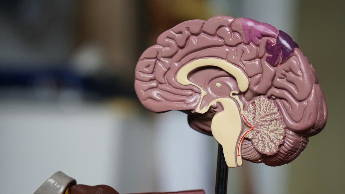
A brain tumour is not the usual explanation for a headache. Most headaches come from migraine, tension, sinus trouble, poor sleep, eyesight, or a short illness. A tumour is uncommon. That fact should steady you, not make you ignore a change that is new, progressive, or paired with another symptom.
This article is general information from a neurosurgical clinic. It does not diagnose you, read your scan, or decide whether you need an operation. If a symptom is sudden or severe, use emergency services. For a planned review, a consultation is the right place to put the story and the reports together.
What a brain tumour is
A tumour is a growth of cells that should not be there, or that are growing in a way the brain did not plan. Some grow slowly and stay in one place. Some grow faster. Some begin in the brain. Some have travelled from a cancer elsewhere in the body. The word covers a wide range, which is why a name on a report is not, by itself, a treatment plan.
The skull is a closed box. A growth, or the swelling around it, can press on brain tissue, on the fluid spaces, or on nerves that leave the brain. Pressure and location explain most of the symptoms people notice. A small growth in a quiet area may be silent for a long time. A small growth beside a speech pathway, the optic nerve, or the fluid pathways can declare itself earlier.
Why the same word covers different problems
Gliomas arise from the supporting cells of the brain. Meningiomas arise from the lining and are often slow. Pituitary growths sit under the brain, near the optic nerves and the hormone system. Metastases are deposits from a cancer that started elsewhere. Acoustic neuromas, also called vestibular schwannomas, arise on the hearing and balance nerve. Cysts and abscesses can mimic a tumour on the story, which is one reason a scan is interpreted by a doctor and not by a headline.
None of these names tells you, on its own, whether to operate next week, to watch, to take medicine, or to treat a cancer elsewhere first. Age, symptoms, the scan, other illnesses, and what you hope to protect all belong in that decision.
A headache that deserves a second look
Headache is common. Tumour is not. The headache that worries a neurosurgeon is usually a change in pattern, not a familiar migraine that has behaved the same way for years.
People describe a new headache that is worse on waking, that wakes them from sleep, or that is joined by vomiting without much nausea beforehand. They describe a headache that is steadily taking more of the week. They describe a headache that has changed character after decades of a different pain. Coughing, bending, or straining may briefly worsen a pressure-type pain. None of these features proves a tumour. Together with other signs, they are a reason to be examined.
A sudden, worst-ever headache is a different problem. It can be a bleed. That is an emergency, not a blog to finish and not a routine clinic slot.
Vomiting, nausea, and appetite
Pressure inside the head can cause vomiting, sometimes with surprisingly little warning. People also lose appetite, feel unsteady in the morning, or notice that tablets for ordinary headache no longer touch the pain. Vomiting has many causes, including infection, migraine, and stomach illness. Persistent morning vomiting with headache, or vomiting with drowsiness or a new weakness, should be assessed promptly.
Seizures in adult life
A first seizure in an adult is a reason for urgent medical care. A tumour is one of several possible causes. Scars, stroke, infection, metabolic illness, and epilepsy that was never recognised can all present this way. The seizure may be a convulsion. It may also be a short spell of unresponsiveness, a strange smell, jerking of one hand, or a wave of déjà vu that is new and repetitive.
Do not drive after a seizure until a doctor has advised you. Do not start or stop seizure medicine on the basis of an article. If a seizure lasts more than a few minutes, or a second seizure follows before recovery, call emergency services.
Weakness, numbness, and one-sided change
The brain organises movement and feeling in a map. A growth that presses on that map can weaken an arm, a leg, or one side of the face. The weakness may be subtle: a cup slips, a foot scuffs, fine work with the fingers becomes clumsy. Numbness, tingling, or a limb that does not feel like your own can appear in the same region.
A sudden weakness, especially with a facial droop or trouble speaking, is treated as a possible stroke until proven otherwise. Call emergency services. Do not wait to see whether a tumour article explains it.
Speech, language, and understanding
Some people cannot find ordinary words. Others speak in sentences that do not hold together. Reading, writing, or understanding what is said can slip. Family members often notice this before the person does. A gradual change over days or weeks needs a medical review. A sudden change is an emergency.
Vision
Vision can fail in several patterns. Peripheral vision may shrink, so people bump into doorframes or miss cars from the side. One eye may blur. Double vision may appear when the eyes no longer line up. A pituitary growth can press the optic nerves and take the outer fields of both eyes. Papilloedema, swelling at the back of the eye from raised pressure, is a finding a doctor looks for. It is not something you can see in a mirror.
Any sudden loss of vision needs same-day care. A slower blur, a new need to sit closer to the television, or double vision that comes and goes should not be left for months.
Balance, coordination, and walking
The cerebellum and the brainstem coordinate movement. Pressure there can cause unsteadiness, a veer to one side, tremor when reaching for a cup, or vomiting with very little headache. People say the world moves, or that they feel as if they are on a boat. Ear disease is a more common cause of spinning vertigo. Persistent imbalance with headache, double vision, or weakness still needs assessment.
Hearing and facial feeling
A growth on the hearing nerve may begin as one-sided hearing loss, tinnitus, or unsteadiness. Facial numbness or pain can come from pressure on the trigeminal nerve. These symptoms also come from ear disease and from trigeminal neuralgia without a tumour. A one-sided hearing change is still worth a proper hearing test and, when the story fits, a scan.
Memory, mood, and personality
Families sometimes describe a person who is less interested, more irritable, unusually sleepy, or no longer able to manage bills and medicines. Frontal and temporal growths can look, at first, like depression, stress, or ageing. A clear change from the person’s own baseline, especially with headache or a seizure, should be examined rather than explained away.
Sleepiness that progresses over hours, confusion, or a person who cannot be woken is an emergency.
Hormones and the pituitary
The pituitary sits below the brain. A growth there may cause headaches and visual field loss. It may also change periods, fertility, thirst, weight, skin, or the ability to tolerate illness. Some people notice milk from the breasts when they are not feeding an infant, or a loss of libido. These symptoms have many hormonal causes. They are part of a medical assessment, not a reason to assume a tumour, and not a reason to ignore a visual change.
Children
Children do not describe symptoms the way adults do. Parents notice a new squint, morning vomiting, a growing head circumference in an infant, a change in school performance, clumsiness, or a seizure. A child who is steadily less playful, or whose headache is waking them, needs a paediatric assessment. This page cannot replace that.
Symptoms that should not wait
Use emergency services for a sudden worst headache, a seizure that does not stop, sudden weakness, sudden trouble speaking, sudden loss of vision, a rapidly falling level of alertness, or a severe head injury. These situations are time-sensitive. A clinic appointment later in the week is the wrong door.
Arrange a prompt medical review, rather than an emergency call, for a new pattern of headache with vomiting, a first short seizure that has fully recovered, gradual one-sided weakness, progressive imbalance, or a clear change in thinking. If you are unsure which door you need, treat the more urgent path as the safer one.
What the examination is for
A consultation starts with the story. When did it begin? What else was happening? Which medicines, including blood thinners and seizure tablets, are already in use? The examination then tests vision, eye movements, facial movement, limbs, sensation, reflexes, balance, and speech, as the story directs. The point is to decide whether the symptoms localise to a place in the nervous system, and how quickly they are moving.
Scans, and what they do not decide
A CT scan is often the first picture in an emergency because it is fast and it shows bleeding and large shifts. An MRI shows softer detail and is the usual scan when a tumour is the question and the person is stable. Contrast dye is used when it will change the reading. Not everyone can have an MRI. Metal implants, some devices, and severe claustrophobia are discussed before the scan is booked.
A scan can suggest a type of growth. It cannot always name it with certainty. It cannot tell you how you will feel after treatment. It should be read with the examination, not instead of it. Bring the actual images if you can, not only the printed report. Reports omit details that the pictures still hold.
A result is not an instruction
People often arrive having read that an operation, radiotherapy, or chemotherapy is “the” treatment for the word on their report. Treatment is chosen for a person. A small, slow meningioma in an older adult with no symptoms may be watched with repeat scans. A growth causing blindness, pressure, or worsening weakness may need to be treated sooner. A metastasis may need the oncology plan and the neurosurgical plan to be written together. Radiotherapy and radiosurgery are options in selected cases. They are not interchangeable with an operation, and they are not suitable for every diagnosis.
Medicine may be used to reduce swelling, to prevent seizures, or to treat a hormone problem. Those medicines have their own risks. Doses are individual. This article will not list them.
What to bring to the visit
Bring every previous scan on a disc or as a file the clinic can open, the written reports, the list of medicines, allergy information, and a short note of when each symptom started. A family member who has watched the change is useful, because the person with the symptoms may not remember the sequence. Write your questions down. In the room, the important ones are easy to forget.
Questions that help
Ask what the scan most likely shows, and what it cannot yet prove. Ask whether waiting is safe, and what change should bring you back sooner. Ask what an operation would be trying to protect, and what it might cost in weakness, vision, or seizure risk. Ask about alternatives, including watchful waiting and non-surgical treatment. Ask who else should be in the discussion, such as an oncologist, an endocrinologist, or an ophthalmologist.
Myths that delay care, and myths that create panic
A normal eye test does not rule out a tumour, and an abnormal eye test does not prove one. A mobile-phone habit has not been shown to be a simple explanation you can use to dismiss a progressive symptom. Yoga, a special diet, or a supplement is not a treatment for a mass inside the skull. At the other extreme, a single ordinary headache after a poor night is not a reason to demand an emergency MRI. The useful middle is a doctor who has heard the timeline.
After a diagnosis
If a tumour is confirmed, the next conversations are about control, symptoms, and function, not about a promise. Some people need an operation. Some need a biopsy so that the treatment can be chosen properly. Some are observed. Rehabilitation, seizure care, and hormone follow-up matter as much as the day of surgery. Recovery is measured in walking, speech, work, and independence, not only in the appearance of a scan.
How this clinic can help
NEUROKALP sees people who need a neurosurgical opinion on brain and spine problems, including when a scan has already raised the possibility of a tumour. The visit is for interpretation and a plan. It is not an emergency department. If you are in the middle of a sudden neurological illness, call emergency services first.
Bring the images. Tell the story in the order it happened. A careful opinion starts there.
Where the growth sits changes the story
The brain is not one organ doing one job. Speech, movement, vision, memory, balance, and hormones live in different neighbourhoods. A symptom is often a clue to the neighbourhood, not a measurement of how large a growth is.
A problem toward the front of the brain may show up as a change in judgement, initiative, or social behaviour. Families say the person is “not himself,” slower to start a task, or newly blunt. Headache may be mild or absent. Weakness, when it comes, often affects the opposite side of the body, because the motor pathways cross.
A problem beside the movement and sensation areas may begin with a clumsy hand, a dragging foot, or numbness that does not follow a single nerve in the arm. A seizure that starts in one thumb and marches up the arm is a classic localising story. It still needs a doctor. It is not, by itself, proof of a tumour.
A problem in the temporal region, at the side of the brain, may affect memory, the naming of objects, or the understanding of speech. Some people notice unusual smells, a rising feeling in the stomach, or short spells in which they are briefly “not there.” The dominant side, usually the left in right-handed people, carries more of the language. The other side carries more of visual memory and the reading of space. These are tendencies, not rules that a person can apply at home.
A problem at the back of the brain may disturb vision even when the eyes themselves are healthy. People miss one side of the page, bump into objects on one side, or lose a portion of the visual field. An eye test that only checks the central chart can miss this. Formal field testing is a different examination.
A problem in the cerebellum produces unsteadiness, a tremor that appears as the hand approaches a target, slurred speech, or vomiting. People are sometimes told they have an ear problem or “vertigo” for weeks. If imbalance is increasing, or if headache and double vision have joined it, the story needs a neurological look.
The brainstem is a crowded passage for nerves that move the eyes and the face, and for pathways that keep a person awake and breathing. Symptoms here can include double vision, facial weakness, swallowing trouble, and long-tract weakness. This is not a region in which to wait and watch a new deficit at home.
A growth in or beside the fluid spaces can block the flow of cerebrospinal fluid. The ventricles enlarge. Pressure rises. Headache, vomiting, sleepiness, and an unsteady walk follow. In an infant the head may grow and the soft spot may bulge. Blocked fluid pathways can become urgent even when the growth itself is slow, because the pressure is the immediate threat.
Raised pressure is a pattern, not a single pain
Raised pressure inside the skull is a clinical picture. Morning headache, vomiting, blurred vision, and a person who is harder to wake are the parts families most often describe. Double vision from a compressed nerve can appear. The examination may show swelling of the optic discs.
Ordinary migraine can also cause severe pain and vomiting. The difference is the company the headache keeps, and whether the person is changing between attacks. A doctor separates these. An article cannot.
Pressure that rises quickly is an emergency. Drowsiness, a new and widening pupil, repeated vomiting, a seizure that continues, or a person who stops making sense over a few hours should go to an emergency department. Do not offer food, do not wait for a relative to finish work, and do not plan to “show the scan at the clinic tomorrow.”
Slow change and fast change are different problems
A meningioma may grow over years. The person adapts, and the family only notices when a small extra demand, such as a festival, a journey, or an illness, makes the deficit obvious. A high-grade tumour, a bleed into a tumour, or a sudden block of fluid can change someone over days. The speed of change often matters more than the name the internet supplies.
Write the timeline down before the visit. “June: harder to find words. August: the right hand tired while writing. September: one seizure.” That sequence is more useful than a stack of adjectives.
Words that appear on a report
Radiology reports use cautious language on purpose. “Suggestive of,” “cannot exclude,” and “clinical correlation is advised” mean the picture has a differential, not a verdict. Enhancement with contrast means the blood–brain barrier in that spot is leaky. It happens with tumours, but also with inflammation, infection, and recent injury. Oedema is swelling in the surrounding brain. Mass effect means something is pushing. Midline shift means the brain is being displaced. Hydrocephalus means the fluid spaces are under pressure. None of these phrases is an instruction to operate that same day, and none of them is a reason to feel reassured without a consultation.
A report that says “likely meningioma” is still a likelihood. The final name, when it is needed, may come from the tissue. Many people are treated on the basis of a very characteristic scan without a rush to surgery. That choice belongs to the person and the surgeon who have seen the images together.
“Benign” is not the same as harmless
Benign, in this context, usually means the cells are not expected to spread through the body, and the growth is slow. A benign tumour can still blind, disable, or threaten life if it sits in a critical place or blocks the fluid pathways. Malignant means the cells behave more aggressively. It does not mean that nothing can be done, and it does not mean that every malignant tumour is treated the same week in the same way.
People carry these words home and hear only the worst sentence. Bring the report to someone who treats the condition. Ask what this word changes in the next month, not only what it means in a textbook.
If the growth began somewhere else
Some brain lesions are metastases, deposits from a cancer of the lung, breast, kidney, skin, or another organ. Sometimes the brain symptom is the first sign, and the search for the source comes afterwards. Sometimes the cancer is already known, and the question is whether the brain finding explains a new weakness or seizure.
The plan is then shared. An oncologist, a neurosurgeon, and often a radiation oncologist look at the number of deposits, the control of the original cancer, the steroid requirement, and what an operation would change. Removing one deposit is sometimes the right step, so that pressure is relieved and the tissue is known. It is not automatically the right step when there are many deposits or when the person’s overall health will not tolerate an operation. This is a judgement, not a slogan.
What an operation is for
Surgery is considered for a reason that can be stated in one or two sentences. Relieve pressure. Restore a blocked fluid pathway. Obtain tissue so the next treatment is not a guess. Remove a growth that is causing a deficit and can be removed at an acceptable risk. Reduce the bulk of a tumour before radiotherapy.
The aim is not always “remove every cell.” Around speech, movement, the brainstem, and the optic nerves, the aim is often the maximum safe removal. Leaving a thin rim beside a working pathway can be the difference between a person who walks and a person who does not. That trade is discussed before the operation, not discovered afterwards as a surprise.
Some operations are biopsies. A small sample is taken, often with image guidance, because the treatment of a lymphoma, an infection, or a particular glioma is completely different. A biopsy has risks of its own, including bleeding and a sample that is not diagnostic. It is chosen when the answer will change care.
What an operation cannot promise
No honest surgeon promises that a tumour will never return, that a seizure will never happen again, or that weakness will fully reverse. Some deficits recover when the pressure comes off. Some are already an injury to the pathway and only partly improve. Swelling after surgery can temporarily worsen a deficit. Infection, bleeding, spinal fluid leak, and new seizures are risks that are quoted for the specific operation, not copied from a general article.
Ask for the risks that apply to this scan and this approach. A percentage taken from a different hospital, a different diagnosis, and a different decade is not your risk.
Radiotherapy, radiosurgery, and medicine
Radiotherapy uses focused radiation to slow or control selected tumours. It may follow an operation, or it may be the main treatment when an operation would not help or would cost too much function. Radiosurgery is a precise, usually single or few-session form used for selected small targets, including some metastases, some vestibular schwannomas, and some arteriovenous malformations. It is not a gentler version of every open operation. The target has to be the right size, the right distance from the optic nerves and brainstem, and the right diagnosis.
Chemotherapy and newer targeted medicines are oncology treatments. They depend on the tissue diagnosis. A neurosurgical article should not list regimens. If those medicines are likely, the oncologist should be in the conversation early.
Steroids are sometimes used to reduce swelling and buy clearer function. They are not a treatment of the tumour, and they have costs: sleep disturbance, blood sugar, infection risk, muscle weakness, and mood change. The dose and the duration are prescribed. They are not adjusted from a blog.
Anti-seizure medicines are used when a seizure has occurred, and in some situations around an operation. They are not started for every headache. Stopping them suddenly is dangerous. Driving rules after a seizure are a legal and medical matter in India as elsewhere. Ask explicitly.
Watching is a treatment, when it is chosen
A small, incidental meningioma found on a scan done for another reason, in a person with no related symptoms, is often observed. The plan is a repeat scan at an interval the surgeon sets, and a clear instruction about which new symptoms should bring the date forward. Watching is not neglect. It is a decision that the risk of treating now is higher than the risk of measuring again.
Watching is the wrong plan when vision is failing, when pressure is rising, when a deficit is progressing, or when the scan looks like something that should not be left unnamed. If you have been advised to wait and you are getting worse, go back. Do not wait for the calendar if the symptom has changed.
Daily life while you wait for a visit
Keep a simple diary: headache, vomiting, seizures, new weakness, and any day you could not do a usual task. Take medicines already prescribed, at the prescribed dose. Do not add painkillers in large combinations, and do not borrow another person’s seizure tablets. Avoid driving if you have had a seizure, unexplained blackouts, or a visual field loss. Ask before air travel if you have recently been told there is swelling or raised pressure. Alcohol and missed sleep make seizures more likely in people who already have that risk.
Tell a family member the plan. If you live alone and a seizure is part of the story, that is relevant to how soon you should be seen and whether you should be staying with someone.
A second opinion
A second opinion is reasonable for a decision about an operation, for a report you do not understand, or when the first advice and your symptoms do not seem to match. Bring the same images. A new opinion made from a verbal description of a scan is weaker than an opinion made from the pictures. It is fair to tell the second surgeon what the first one advised. The goal is clarity, not a collection of contradictory sentences.
What to ask, written so you can take it in
Ask where the growth is, and which function lives next to it. Ask what the report is confident about, and what still needs tissue or time to prove. Ask what is likely to happen over the next month if nothing is done. Ask what would count as worsening. Ask whether the proposed operation is for removal, for pressure, or for diagnosis. Ask what function is being placed at risk on purpose, because it sits in the way. Ask who else should see you before a decision: oncology, endocrinology, ophthalmology, or radiation oncology. Ask about the practical recovery: walking, speech, wound care, and when work is usually discussed. Ask what the clinic will do if you deteriorate before the planned date.
Myths that waste time
A normal blood test does not clear the brain. A CT done years ago does not describe the head you have today. Pain that responds to a tablet can still be serious, and pain that does not respond is not automatically a tumour. Glasses do not treat a field defect from a pituitary mass. Ear drops do not treat a one-sided hearing loss from a nerve sheath tumour. A diet, a juice, or an alkaline claim will not shrink a mass inside the skull. Refusing a scan because of fear of the machine delays the only information that can settle the question.
Fear of the word “tumour” also causes harm in the other direction. People demand emergency surgery for a finding that is safe to discuss next week. Urgency is a medical judgement based on pressure, deficit, and speed. It is not a feeling that the waiting room is taking too long, and it is not a feeling that can be ignored when the person is becoming drowsy.
After a decision, the work continues
If an operation is planned, the useful preparation is practical. Sort medicines, especially blood thinners, only under instruction. Arrange a companion for the admission. Know which symptoms before the date should cancel the wait and send you to emergency care. After surgery, the early goals are safe wakefulness, a wound that is dry, pain that is controlled, and a clear instruction for steroids or seizure tablets if they were prescribed. Rehabilitation starts early when strength, speech, or balance have been affected. Follow-up imaging is timed to the diagnosis, not to anxiety alone, though new symptoms always outrank the calendar.
If radiotherapy is planned, the conversation shifts to timing, hair and skin effects, fatigue, and the weeks over which swelling can temporarily increase. If observation is planned, put the next scan date in writing and the warning symptoms beside it.
Questions people ask
Is every brain tumour cancer?
No. Many tumours that arise from the coverings of the brain, the nerve sheaths, or the pituitary are not cancers in the sense of a disease that spreads through the body. Some are. The word on the report has to be explained for that diagnosis.
Can a tumour be present with no headache?
Yes. Headache is not required. Seizures, a quiet change in behaviour, a field defect, or one-sided hearing loss can be the first sign.
Does a long history mean it is safe?
A long, stable history is often reassuring, and it fits many slow growths. A long history that has begun to accelerate is a different story. The recent change matters.
Should I get an MRI for every new headache?
No. Most headaches do not need imaging. Imaging is considered when the examination is abnormal, the pattern is atypical, the headache is progressive, or there are seizures, cancer, immune problems, or neurological signs. A clinician decides.
Can stress or a mobile phone cause these symptoms?
Stress changes sleep and pain. It does not explain a progressing weakness, a seizure, or a field defect. Mobile-phone use is not a reason to skip a scan that has been advised for a progressive symptom.
What if two doctors disagree?
Ask them what fact they are disagreeing about: the name, the speed, or the risk of operating. Often the disagreement is about timing, and a short period of planned review resolves it. If the disagreement is about whether the pressure is dangerous, do not leave that unresolved over a weekend.
Can I exercise?
Walking, if you are steady and have not been told to rest, is usually reasonable. Do not swim alone, drive, or work at heights if seizures or blackouts are part of the story. Ask about the specific restriction rather than stopping every activity out of fear.
Will I be awake during surgery?
Only in selected operations, usually when a language or movement pathway must be tested during the removal. Most brain operations are done under general anaesthesia. Awake surgery is a technique for a particular problem, not a standard you should expect or fear in advance of a consultation.
A last distinction
Learn the warning signs so that you do not ignore them. Do not use the warning signs to diagnose yourself. The consultation exists to hold the story, the examination, and the images in one place, and to say what should happen next. That is the work of the visit.
How a first consultation is usually built
The visit begins with the timeline, not with the scan. A surgeon wants the first day you noticed something, what you were doing, whether it came and went, and what has changed in the last fortnight. Medicines matter: blood thinners, seizure tablets, steroids already started elsewhere, and any drug you buy yourself for pain. Other illnesses matter: a known cancer, a clotting disorder, a previous head injury, tuberculosis treatment, or a problem with the immune system. These change what a white patch on a scan is likely to be.
The examination is a series of small tests. Vision, fields, pupils, and eye movements. Face, hearing, palate, and tongue. Arms and legs for power, tone, reflex, and feeling. Walking, if it is safe. Speech and a few questions of memory and attention. None of this is a formality. A scan that looks worrying and an examination that is normal lead to a different conversation from a scan that looks quiet and an examination that shows a new field cut.
Only then do the images take the centre of the table. Looking at them together is part of the explanation. You should leave knowing where the finding is, what sits next to it, and what the next step is. If you leave with only a printed adjective, ask for the sentence that was missing.
Preparing for an MRI or a CT
An MRI is noisy and still. You lie in a tunnel while the machine takes a long set of pictures. Tell the department about pacemakers, cochlear implants, aneurysm clips, infusion pumps, and metal in the eyes from old industrial work. Most modern joint replacements are safe, but the department checks the exact device. Contrast dye is used when it separates one diagnosis from another. Mention kidney disease and previous reactions to dye before the injection, not afterwards.
A CT is faster and is the usual first picture when someone is drowsy, has just had a seizure, or may have a bleed. It uses X-rays. It is less sensitive for many small or soft tumours, so a normal CT does not always close the question. If the story is progressive, an MRI is often the next picture, not a reason to argue with the first doctor who correctly started with CT in the emergency room.
Claustrophobia is common. Say so when the scan is booked. A short-acting sedative is sometimes used, and you will then need someone to take you home. Children, and some adults, need an anaesthetic for a still scan. That is arranged, not improvised.
Bring the disc. A report without pictures forces the next doctor to repeat the scan or to guess. If the disc will not open, ask the centre for a copy in a standard format before you travel.
Findings that mimic a tumour
Not every mass is a neoplasm. An abscess can look rounded and can cause headache, fever, and a seizure. In India, tuberculoma and neurocysticercosis are part of the differential for a ring-enhancing lesion, especially when the person is young and the illness has been subacute. Inflammation, demyelination, and a recent stroke can also occupy a place on the list. This is why a surgeon sometimes asks for blood tests, a chest image, or a short period of treatment and a repeat scan before anyone talks about an operation.
If you have been told “it might be infection or it might be a tumour,” that is an honest sentence. The way to resolve it is a plan: which test, by when, and what symptom would make the plan urgent. It is not a reason to collect opinions until the pictures are forgotten.
Pituitary and skull-base problems in more detail
The pituitary sits in a bony hollow beneath the brain, between the optic nerves. A growth here may press upward and take the outer half of the visual field in each eye, so that people still read a chart with the centre of their sight and still collide with vehicles from the side. Hormonal change can be the louder symptom: periods stopping, milk from the breasts, loss of libido, unusual thirst and large volumes of urine, weight change, or a failure to recover from an ordinary illness because cortisol is low.
Some of these growths are treated with medicine first, particularly many prolactin-secreting adenomas. An operation, often through the nose with an ear, nose, and throat colleague, is considered when vision is threatened, when medicine has not worked, or when the type of growth is not one that medicine shrinks. This is a combined clinic problem. A neurosurgeon, an endocrinologist, and an ophthalmologist are often all needed. Starting or stopping hormone tablets without that team is unsafe.
Skull-base growths, including vestibular schwannomas, sit deep beside the hearing and balance nerves and the facial nerve. One-sided hearing loss and tinnitus are the usual opening. Balance follows. Facial weakness is a later and more worrying sign. Treatment may be observation, radiosurgery, or an operation, chosen according to size, hearing that remains, growth on serial scans, and age. Rushing a small, stable schwannoma into surgery is not automatically the careful choice. Ignoring a growing one because the word “benign” appeared on the first report is not careful either.
Seizures, work, and the road
A single seizure deserves assessment the same day if it is the first, and emergency care if it is prolonged or repeated. Afterward, the practical questions arrive quickly. Can I ride a two-wheeler? Can I drive the family car? Can I return to a job on a ladder, with machinery, or with children in my sole charge? The safe answer is specific to the type of spell, the scan, and the medicine, and it should be written down. Until that advice exists, do not drive and do not work at height.
Witnesses are valuable. Ask the person who saw the event what the eyes did, whether the body was stiff or jerking, how long it lasted, and how long it took for speech to return. A video, taken only when the person is safe and not restrained roughly, can be more useful than a long description. Do not put anything in the mouth.
Older adults, and younger adults
In an older person a short story of confusion is easily blamed on age, a urine infection, or a sleeping tablet. Those are real causes. A new one-sided weakness, a new seizure, or a headache unlike any previous pain still needs an examination. Treatment decisions then weigh fitness for anaesthesia, bone health, and what the person wants to preserve: living at home, speech, the ability to recognise family.
In a younger adult the same symptoms are sometimes dismissed as stress, screens, or a heavy week. Progressive deficits are not a lifestyle problem. At the same time, a young person with a single brief symptom and a normal examination may need reassurance and a plan rather than an immediate operation. Age changes the probabilities. It does not replace the examination.
Pregnancy
Headache in pregnancy is usually migraine, blood pressure, or the ordinary discomfort of the months. New seizures, a persisting visual change, or a progressive weakness are not ordinary. Scans can be planned with obstetrics so that the mother and the pregnancy are both considered. Medicines, including seizure tablets and any steroid, are chosen with that same joint view. This page will not rank those medicines. The emergency rule does not change: a sudden neurological collapse is an emergency in pregnancy as it is at any other time.
What families can usefully watch
Families often see the change first. The useful report is concrete. She missed the salt on the left side of the plate. He stopped midway through a sentence and did not notice. The right foot caught on the step three days running. Vomiting on waking, three mornings. A vague “she is not normal” is a start, but the example is what the doctor can use.
Agree on one person to speak if the patient is tired. Bring the medicine boxes, not a recollection of the doses. Decide before you leave the clinic who will watch for the warning symptoms that night, and which hospital you will use if drowsiness or a seizure appears. Hope is compatible with a plan.
A simple note to keep before the visit
Write five lines.
- The first symptom, and the month it began
- The symptom that has changed in the last two weeks
- Any seizure, blackout, or fall, and whether someone saw it
- Current tablets, including blood thinners and steroids
- The question you most want answered in the room
That note keeps the visit on the decision, which is where it belongs.
More questions people ask
Do I need a scan before I book a neurosurgical visit?
Not always. If the symptoms are new and progressive, a physician or a neurologist may arrange the right scan first. If a scan already exists, bring it. A visit without images can still be useful for the examination and for deciding which scan, if any, is next. A visit is not the place for a sudden collapse. That is the emergency department.
The report says oedema. Does that mean the tumour is large?
Oedema means swelling in the brain around a lesion. A small lesion can have a large amount of swelling, and that swelling can cause more of the symptoms than the mass itself. Swelling can be treated while the diagnosis is clarified. It is not a number you can compare with another person’s report.
Can physiotherapy cure a brain tumour?
Physiotherapy helps weakness, balance, and the return to walking. It does not remove a mass. It is part of recovery, and it is sometimes part of the waiting period so that a person stays as fit as possible. It is not an alternative to a scan that has been advised.
Should the whole family be told?
Tell the people who will notice a change and who will have to act. Children usually do better with a simple true sentence than with a silence they can feel. The sentence can be short: there is a problem in the brain, doctors are finding out what it is, and this is who to call if someone becomes very sleepy. Detailed prognosis talks belong with the doctors who have the images, not in a hallway guess.
Is it safe to wait for a festival, a wedding, or a journey?
Sometimes a date can move, and sometimes it must not. Ask directly: “If we wait three weeks, what are we risking?” If the answer is vision, pressure, or a progressing deficit, the celebration can be rearranged. If the answer is that the finding is stable and the wait is already the plan, then the wait should be explicit, with a scan date and warning signs.
What does follow-up look like if we do operate?
Wound review, a check of the function that was at risk, a plan for steroids or seizure tablets if they were used, and an imaging date that matches the diagnosis. Some people need radiotherapy planning in the same month. Some need only a scan months later. The schedule is part of the consent conversation, not an afterthought.
What if I cannot afford to repeat the MRI immediately?
Say so. A clinician can often judge whether the existing images are enough for the next decision, whether a CT will answer the urgent question, or whether a short delay is medically acceptable. Pretending the disc will arrive, and then disappearing, is more dangerous than an honest conversation about cost and timing.
What this page will not do
It will not tell you that your headache is a tumour. It will not tell you that your headache is harmless. It will not name an operation, a dose, or a hospital package. Those are decisions for a doctor who has examined you and seen your images. Use the page to recognise a change, to know which changes are emergencies, and to walk into a consultation with a clear timeline and a clear question.
Between the visit and the next scan
Waiting is easier when the wait has a shape. Write down the date of the next image, the name of the person to call if a symptom changes, and the hospital you will use in an emergency. Keep the disc or the file with your identity papers so a night-time doctor is not starting from a blank screen.
A stable finding can still produce a bad week. Poor sleep, a viral illness, or a missed meal can worsen headache in anyone. The change that should move the date forward is a new kind of symptom: a first seizure, a new weakness, vomiting that has become morning vomiting, a visual change, or a person who is harder to wake. Familiar pain that is a little louder for a day, in a person who is otherwise themselves, can often wait for the planned review. If you are unsure, treat it as the more urgent of the two and ask.
Do not start a steroid, a seizure tablet, or a sleeping medicine because a relative was given one. Those drugs are matched to a person. Do not stop a prescribed steroid suddenly. The dose is tapered by the doctor who started it, because an abrupt stop can make a person seriously unwell.
How to read the next report without frightening yourself
Compare the new report with the old one only on the points your surgeon marked: size, swelling, shift of the midline, and the fluid spaces. A different radiologist will use different adjectives for the same picture. “No significant interval change” is often the sentence that matters. A new sentence about enhancement, bleed, or hydrocephalus is a sentence to ask about promptly, not at the next festival.
Measurements in millimetres move with the angle of the slice. A one-millimetre difference between two centres is not automatically growth. Growth is a judgement made by someone looking at both studies, not a subtraction you do in the corridor.
Work, study, and the people who depend on you
Ask for written limits rather than guessing them. Some people can do desk work while a small, stable finding is observed. Some should not drive, climb, or be the only adult with a child if seizures are part of the story. Employers usually need a short note of fitness and review date, not the entire report. You can ask the clinic what may be shared.
Students sitting examinations deserve the same honesty. Extra time, a nearby washroom, or a deferred paper is a conversation with the institution. It is not a reason to hide a progressing symptom until the examination is over.
If the diagnosis is still uncertain
Uncertainty is a medical state, not a failure of the visit. Some scans need tissue. Some need a short interval and a second picture. Some need a physician to look for infection or a cancer elsewhere before a neurosurgeon can give a useful opinion. Ask what would make the uncertain period unsafe. Then live inside the boundary you were given, and come back if the boundary is crossed.
Bring a family member to the visit where the name is first explained. People remember different halves of the same conversation. A written line from the surgeon, even three sentences, prevents a week of arguing about what was said.
Care that sits beside the operation
Nutrition, sleep, and treatment of blood pressure or diabetes do not shrink a tumour. They do change how safely a person can undergo an anaesthetic, heal a wound, and join rehabilitation. Mention every illness, including ones that feel unrelated. Bring the latest sugar readings or blood-pressure notes if you have them.
Mood changes are common when a person is waiting on a brain scan. Irritability and poor sleep are not proof that the growth has reached the frontal lobe, and they are not something to be ashamed of. Tell the doctor if worry has become panic, if you have stopped eating, or if you feel unsafe. That is part of care.
A clear close
A brain tumour is an uncommon explanation for everyday headache, and a serious explanation for a progressing neurological change. Learn the difference between a familiar pain and a new pattern. Use emergency services for a sudden collapse, a prolonged seizure, a sudden loss of speech, strength, or vision, or a person who cannot be woken. Use a planned consultation to put a slower story next to the images.
NEUROKALP offers that planned opinion for brain and spine problems. Bring the pictures, the medicine list, and the five-line timeline. The visit is for a decision about what should happen next. It is not a substitute for emergency care, and it is not a promise about the outcome. The promise a clinic can keep is a careful reading of your own case.
Symptoms, named one by one
A change in smell, either a loss or a strange odour that no one else notices, can come from the nose, from a virus, or from the pathways under the frontal lobes. It is rarely the only sign of a tumour. Together with personality change or headache, it belongs in the history.
A ringing in one ear, or a phone that is clear on one side and dull on the other, deserves a hearing test. Wax and age explain many cases. A one-sided loss that a test confirms should not stop at wax if the examination of the ear is normal. The hearing nerve is one of the structures a scan is asked to see.
Double vision that is new may be two images side by side or one above the other, and it may vanish when either eye is covered. That pattern suggests the eyes are not aligned, which can be a nerve, a muscle, or pressure inside the head. Covering one eye as a temporary comfort is reasonable on the way to hospital. It is not a treatment.
A droop of one side of the face, with a weak arm or slurred speech, is treated as a stroke until a scan says otherwise. A facial droop with ear pain and a rash is a different illness. Call emergency services for the sudden version. Do not decide between them from a photograph.
Morning vomiting that is forceful, especially in a person who then feels briefly better, is the pattern families describe when pressure is high. Vomiting with diarrhoea and a fever in the whole household is usually infection. The company the vomiting keeps is the clue. Headache, drowsiness, or a new squint in a child moves it out of the “stomach bug” category.
Seizures that start with a turning of the head, a jerk of one hand, or a strange feeling and then become a convulsion are focal seizures with spread. Seizures in sleep, a bitten tongue, and a long confusion afterwards are details worth telling. A faint at the sight of blood, with a pale face and a quick recovery, is often not a seizure. When you are unsure, seek a same-day assessment rather than a label from this paragraph.
Memory loss that is slow and covers recent events equally, without headache, weakness, or a seizure, is often the territory of a physician or a neurologist rather than an urgent tumour search. Memory loss with a one-sided sign, a seizure, or a clear change over weeks is a different tempo. Families should describe examples: unpaid bills, a lost route home, a story repeated every few minutes. Adjectives are less useful than the example.
How to live with a scan that is not yet explained
Sleep in the same house as someone who knows the warning signs if a seizure or raised pressure is already part of the story. Keep a phone charged. Avoid driving. Take the medicines that were started, at the dose that was written. Do not add aspirin because a headache “might be pressure.” Aspirin is not a treatment for a mass, and it complicates any operation that may be needed.
Alcohol lowers the threshold for a seizure and muddles the morning assessment of drowsiness. This is a poor week for it. Heavy exercise, a head-down posture, and a long hot journey are worth asking about if you have been told the pressure is high. Ordinary walking, if you are steady, is usually a kindness to your mood and your sleep.
If a seizure happens while you are waiting, note the time it started and ended, keep the person on their side, and do not put a spoon in the mouth. Call emergency services if it lasts more than a few minutes, if a second seizure follows, if there is an injury, or if breathing is poor. A first seizure is a same-day medical event even if it stops.
Talking about the result
Use the name the doctor used, and ask for a sentence you can repeat. “There is a growth on the covering of the brain. It is slow. We are repeating the scan in four months unless your vision changes.” That kind of sentence prevents a relative from translating “meningioma” into a story the doctor did not tell. If you did not hear a sentence that clear, ask for it before you leave.
Decide who will receive the detailed news. One accurate account is kinder than five half-heard ones. You may ask the clinic what can be written down. You are not obliged to post a scan on a family group with a request for opinions. Those opinions will not have examined you.
Fear of the word does not mean you must decide the operation on the day you hear it, unless the pressure or the vision says the day is exactly that urgent. It is reasonable to say you will return with your questions. It is not reasonable to vanish if you were told the situation can worsen quickly. Leave a phone number either way.
What belongs in a follow-up visit
Bring the new images and the old ones. Bring a note of any seizure, any steroid dose and who changed it, and any new tablet started by another doctor. Mention wound redness, fever, or a leak of clear fluid from the nose after a skull-base or pituitary operation. Mention thirst and very large volumes of urine. Mention a field of vision that seems narrower than at the last visit.
The useful outcome of the visit is a written next step: watch, operate, refer to oncology or endocrinology, or go to emergency care if a named symptom appears. A visit that ends only in reassurance, with no sentence about what would cancel the reassurance, is a visit to complete before you go home.
One page you can keep
Most headaches are not tumours. A new pattern, a seizure, a one-sided weakness, a change in speech or vision, or a person who is harder to wake, is not an ordinary headache. Sudden versions of those signs are emergencies. Slower versions deserve a planned examination and, when it will change the decision, a scan.
Bring the timeline and the pictures. Ask what the finding threatens, what waiting risks, and what an operation would be for. NEUROKALP is a place for that planned conversation. The emergency department is the place for a stroke, a prolonged seizure, a sudden collapse, or a person you cannot wake. Knowing which door to use is the practical point of everything written above.
Words on a brain scan
Mass lesion. A space-occupying finding. It may be a tumour, an abscess, or inflammation. The word does not name the treatment.
Oedema. Swelling in the surrounding brain. It can cause more of the symptoms than the mass itself.
Mass effect. Pushing of nearby structures. It is a description of pressure, not a size grade you can compare on the internet.
Midline shift. The brain has been pushed off the centre line. Significant shift is a reason for a prompt surgical look.
Enhancement. The lesion takes up contrast. Tumours, infections, and inflammation can all do this.
Necrosis. An area inside a lesion that looks dead. It changes the differential. It does not, alone, set the day of surgery.
Hydrocephalus. The fluid spaces are enlarged under pressure. It can be urgent even when the growth is slow.
Midline shift absent, no hydrocephalus. Often the phrase that means the pressure, for the moment, is not the emergency. The diagnosis can still need a plan.
Vasogenic oedema. Swelling that follows a leaky blood–brain barrier, typical around many tumours and abscesses.
Restricted diffusion. An MRI pattern that can suggest abscess or a dense cellular tumour, among other things. It is interpreted with the rest of the picture.
Extra-axial. Outside the brain substance, often from the coverings. Meningiomas are the familiar example.
Intra-axial. Inside the brain substance.
Dural tail. A thickening of the covering beside a mass, often seen with meningioma, not proof by itself.
Optic chiasm compression. Pressure on the crossing of the optic nerves. It threatens the side vision of both eyes.
Pituitary macroadenoma. A pituitary growth large enough to be seen as a mass, often the ones that touch the visual pathways.
Metastasis. A deposit from a cancer that began elsewhere. One deposit and many deposits lead to different plans.
Gliosis. A scar-like change in brain tissue. It is not a tumour, though it can sit beside one on a report that is describing the edge.
The night before a planned scan or a visit
Lay out the disc, the old reports, the medicine boxes, and the five-line timeline. Charge the phone. Eat and drink as the scanning centre instructed. If you were told to fast for contrast or for a possible anaesthetic, follow that instruction rather than a general habit. Take blood-pressure and seizure medicines unless you were told to hold them. Arrive with a companion if you may be sedated or if a seizure is part of the recent story.
Write the one question you will ask if the conversation runs away from you. “What happens if we do nothing for one month?” is often the question that organises all the others. Ask it before you ask about cost, about a wedding date, or about a technology you read of the night before. Those matter. They matter after the risk of waiting is clear.
If, on the morning of a planned visit, the person is drowsy, has had a seizure that will not stay in the past, cannot use one side, or cannot see, do not keep the appointment as if it were an ordinary booking. Go to emergency care. The images can be seen there. A waiting room is the wrong place for a changing brain.
A short list to carry
Keep the emergency numbers, the name of the hospital you would use at night, and the clinic number in the same note on your phone.
Keep the medicine list in the same note, including steroids and seizure tablets, and the date any dose was last changed.
Keep the date of the next scan, and beside it the symptoms that should bring that date forward: a seizure, new weakness, new trouble with speech or vision, repeated vomiting, or increasing sleepiness.
Keep the discs. A report is a summary. The pictures are the evidence the next doctor will trust.
Most days with a stable finding are ordinary days. Live them. The list above is what keeps an ordinary day from becoming a dangerous one without anyone noticing.
If a sign on that list appears, do not reread this article looking for a softer interpretation. Use the emergency door for a sudden or worsening change, and use the clinic for a planned reading of your own images. The right door, used on time, is the whole of the advice.
A careful opinion starts with your timeline and your pictures. Bring both.
This page is for general awareness. It is not a diagnosis or a personal treatment plan. Book a consultation for advice about your own care.